Bioinformatics: Protein Structure
The example gene we studied yesterday "Thioredoxin H" is involved in redox reactions and similar proteins are present in virtually all organisms. What is perhaps interesting and rare about the protein that this gene encodes is that there is X-ray crystal structure for it and for a mutant.
Before we can compare these structures, let's take a moment to learn different persepectives on structure:
Primary Structure: The sequence of amino acids that makes up a protein.
Secondary Structure: The regions of regular substructures such as alpha-helix and beta-sheets.
Tertiary Structure: The 3-dimensional structure of the protein when folded.
Examples:
1. Primary Structure Example: Just a sequence of amino acids like we've seen before

In this example, software has predicted where different structures take place. This can be done by recognizing patterns in the sequence. What are these structures anyway? Alpha Helices and beta sheets are structures formed by hydrogen bonds between amino acids.

As you can see, the precise sequence of amino acids would be important for making these structures.
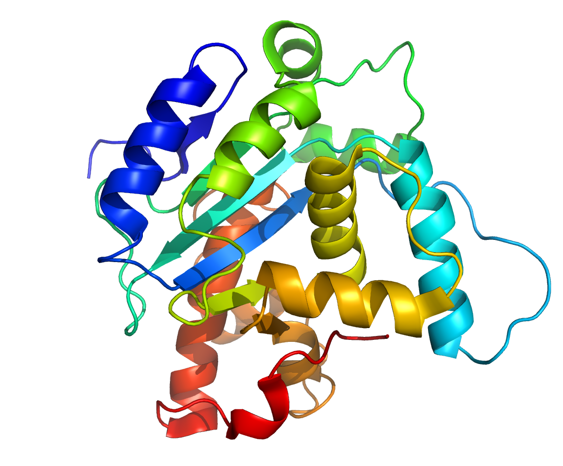
3. Tertiary Structure Example:
 You can see how these alpha helices and beta strands fit into the big picutre of the 3D structure.
Now let's examine the protein structure:
Wild-type Trxh Structure
And the mutant structure:
Mutant Trxh Structure
What differences do you find?
Before we can compare these structures, let's take a moment to learn different persepectives on structure:
Primary Structure: The sequence of amino acids that makes up a protein.
Secondary Structure: The regions of regular substructures such as alpha-helix and beta-sheets.
Tertiary Structure: The 3-dimensional structure of the protein when folded.
Examples:
1. Primary Structure Example: Just a sequence of amino acids like we've seen before
MPILQDSKLVA...2. Secondary Structure Example:

In this example, software has predicted where different structures take place. This can be done by recognizing patterns in the sequence. What are these structures anyway? Alpha Helices and beta sheets are structures formed by hydrogen bonds between amino acids.

As you can see, the precise sequence of amino acids would be important for making these structures.
3. Tertiary Structure Example:
 You can see how these alpha helices and beta strands fit into the big picutre of the 3D structure.
Now let's examine the protein structure:
Wild-type Trxh Structure
And the mutant structure:
Mutant Trxh Structure
What differences do you find?